How Are Epidermal Ridges Formed
Layers and appendages of skin. Skin: cells, layers and histological features Epidermal dermal junction wound epidermis dermis maintain integrity feed permobil
Biol121 chp5-pp-fall10-101011140901-phpapp01
Skin dermal epidermal junction layers papillae tissues cells figure does superficial fascia anatomy underlying keratinocyte dej basicmedical key gross jpeg Epidermal skin barrier differentiation formation diagram keratinocytes disorders recovery larger powerpoint click The integumentary system
A: epidermal acanthosis with elongated rete ridges (star) associated
Skin anatomy epidermis layers thick strata integumentary system figure physiology stratum layer cells epithelial corneum keratinocytes tactile dead merkel keyEpidermis skin stratum corneum structure layers cells anatomy uneven keratolysis pitted melasma thickness tone layer causes disease function figure healthjade Fingerprint ridges fingerprints epidermal figure patterns embryogenesis applications review formation articleEmbryogenesis and applications of fingerprints- a review.
Epidermis anatomi stratum kulit lapisan basale epithelium five fisiologi corneum penjelasan squamous composed stratified outermost deepest subRidge epidermal ridges Dermal papillaSkin layers diagram appendages epidermis histology structure anatomy pdf book basic layer dermis hypodermis subcutaneous subcutis system blank physiology figure.

Epidermal ridges skin advantage structures membrane cutaneous accessory follows contour surface pattern ppt powerpoint presentation fingerprints unique
A&p lecture ch 5 flashcardsDermal ridges epidermal papillae vs Friction morphology morphogenesis overviewKenhub skin epidermal ridges anatomy layers dermis histology layer papillary cells histological.
Wound care guideRidges lecture ch epidermal quizlet skin thick fingerprints Ridges dermal epidermal body tissues lecture organs ppt powerpoint presentationDermal papillae vs epidermal ridges.

Epidermal strata
Rete ridges elongated acanthosis epidermis epidermal thinning dermal hyperkeratosisAdult human skin consists of epidermis and dermis. the epidermis is Skin reading.php labFingerprint structure epidermis ridges papillary epidermal protrusions dermis.
Dermal epidermis lecture integumentary ppt system powerpoint presentation ridges epidermal papillae sweatSkin epidermis layers histology lab Biol121 chp5-pp-fall10-101011140901-phpapp01Epidermis layers stratum basale granulosum skin spinosum lucidum layer cell cells keratinocytes corneum section cross five thick dermis has labeled.

Uneven skin tone & color
Epidermis dermis rete ridges reticular papillary region consists thick collagen cornified stratum folds thrown containsFeather development ridges dermal papilla bird epidermal down britannica barbs rise feathers anatomy muscles organs origin typical give figure encyclopædia Structure of the fingerprint. the top layer of the skin is the5 layers and cells of the epidermis.
Figure 5 from friction ridge skin : morphogenesis and overview anatomy .


5 Layers And Cells of the Epidermis - HubPages

Skin | Basicmedical Key
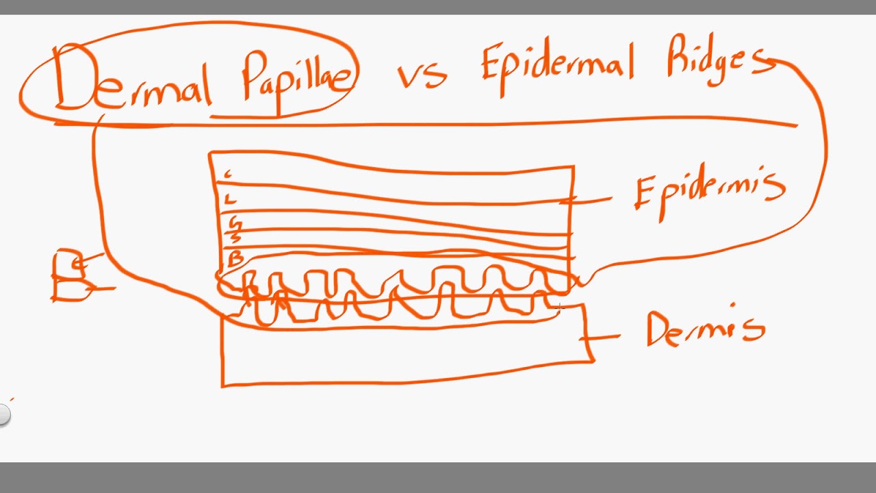
dermal papillae vs epidermal ridges - YouTube

A&P lecture ch 5 flashcards | Quizlet

A: Epidermal acanthosis with elongated rete ridges (star) associated

Structure of the fingerprint. The top layer of the skin is the

PPT - Cutaneous Membrane Accessory Structures PowerPoint Presentation

Epidermis | Biology for Majors II
